3D CNS Cell Interaction Mapping
As part of a research effort investigating immune–glial interactions in neuroinflammatory disease, I imaged CNS tissue from diseased mice and reconstructed each dataset in 3D. The goal was to quantify how specific cell populations interact throughout the injured brain and spinal cord, and how these relationships change during demyelination and recovery. This work complemented broader studies on how oligodendroglia communicate with immune cells during MS-like pathology
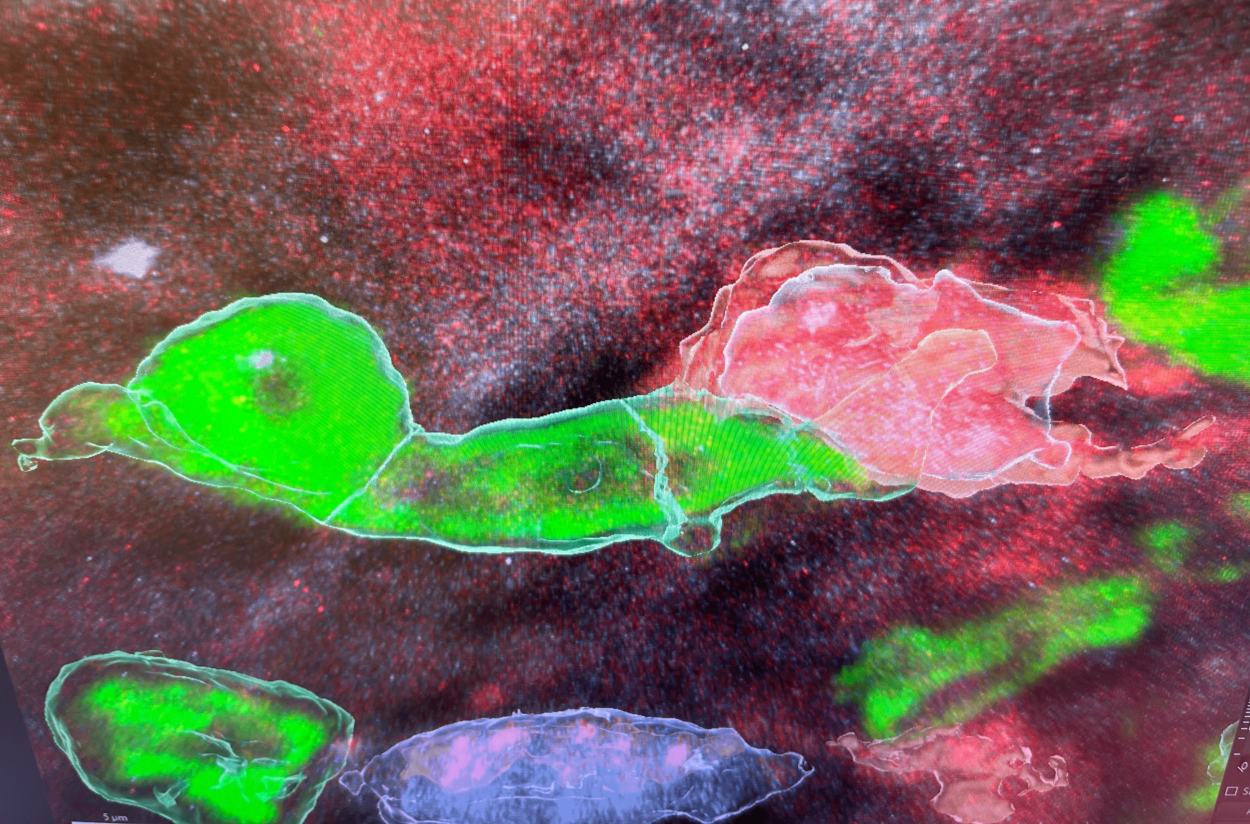
The workflow began with high-resolution confocal imaging of diseased mouse CNS tissue. Each image stack was registered and reconstructed in 3D, allowing individual cells to be segmented and spatially localized. From these reconstructions, I extracted features describing pairwise cellular distances, neighborhood composition, and structural organization across the tissue. These spatial datasets were then used to train machine-learning models that characterized interactions between immune cells (e.g., T cells) and oligodendroglia within demyelinating lesions. The resulting analyses helped identify patterns of cell proximity and organization that may relate to inflammatory activity, remyelination potential, and recovery dynamics, supporting work showing that MHC-expressing oligodendroglia directly engage immune cells in disease contexts.
This work was done in Dr. Peter Calabresi's Lab where I was mentored by Dr. Jingwen Hu